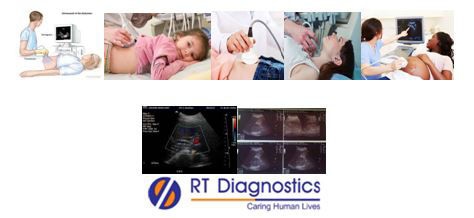
Ultrasound Abdomen:
Why Ultrasound Abdomen Test?
CLINICAL INFORMATION
Diagnostic ultrasound also known as sonography(or diagnostic medical sonography) is one of the imaging studies. For diagnostic use Ultrasound generally uses between 2 to 20 kilohertz or megahertz. Three types of Doppler ultrasound include colour Doppler, power Doppler, and spectral Doppler. It is also known as a surgeon’s guide that helps in their movements during surgical procedures. This technique is based on the application of using high-frequency sound waves to view inside the body (trans-oesophagal echocardiogram, trans-rectal ultrasound, transvaginal ultrasound – gynaecology ultrasound etc), thus helping in the diagnosis and treating a variety of diseases. This technology of ultrasound captures the real-time images. It exhibits movement of the body’s internal organs as well as blow flow through the blood vessels. The advantage of ultrasound over X-Ray imaging is that no ionizing radiation exposure is involved here. A thin layer of water-based gel is applied to the skin and a probe is placed over the area of interest (suspected organ). The application of gel secures contact with the body. These gel, apart from lubrication (prevent friction) acts as an interface and also prevents from the creation of air pockets in the organ. An ultrasound examination uses the application of a probe called a transducer, which is placed directly on the skin and/or inside a body opening. This transducer (probe) transmits ultrasound waves. The ultrasound image depends on the strength (amplitude) of the sound signal and frequency (pitch), along with the time taken for the wave to travel through the body. Hence this ultrasound imaging is a medical tool that deploys technology that helps physicians to evaluate, diagnose, treat medical pathologies (prognosis) etc. Thus an abdominal ultrasound helps the physician to evaluate the cause of abdominal pain or bloating. It also helps to evaluate for angioplasty (vascular stenting), carotid duplex ultrasonography, presence of blockages of blood flow (clots), narrowing of vessels (stenosis), tumours and congenital vascular malformations, urethral stricture, varicocele, bladder dysfunction, diverticula, ischemia (reduced blood flow to organs such as testes, ovaries etc), infection (increased blood flow), frozen shoulder, tennis elbow, carpal tunnel syndrome, identifying ectopic pregnancy etc. The other uses of ultrasound imaging include examining body’s internal organ like liver, spleen, uterus, ovaries, scrotum, structures of infants such as brain, hip, spine etc, bone sonometry (evaluate metabolic bone disease), breast ultrasound, assessment of joint inflammation (synovitis), diagnose gall bladder disease (eg. calculi), Doppler ultrasound to monitor vascular abnormalities, Obstetric ultrasound – Doppler fetal heart rate monitors (pregnancy), Nuchal translucency (sonographic appearance of a collection of fluid under the skin behind fetal neck) and also checks for Down’s syndrome, Echocardiogram, DVT, aneurysm, ophthalmic ultrasound (ocular structures), evaluation of thyroid, detect genital and prostate problems, urinary bladder and kidney stones, evaluation of ascites (pancreatitis, ultrasound-guided biopsies (FNAC, Tumor Biopsy etc), ultrasound-guided needle placement etc. Indications of ultrasound include a diagnosis for the identification to know the cause of the pain, swelling, infection in the body’s internal organ, biopsy such as FNAC (guided by ultrasound), calculus (stones), evaluate hip, spine etc, examine a fetus in a pregnant woman etc. Ultrasound is safe, non-invasive and does not use radiation. Ultrasound technique has many advantages over X-Ray images eg. Ultrasound is devoid of harmful ionizing radiations, unlike X-Ray studies. Moreover, four-dimensional (4D) ultrasound reads and records motions. Demerits of ultrasound include a few pitfalls, including heat generation of tissues and the disadvantage of causing small pockets of gas in body fluids or tissues (cavitation). Since the ultrasound doesn’t travel well in gas (gets disrupted) and hence imaging body parts with gas like lungs are ineffective. Furthermore, expectant mothers should be aware of concerns with purchasing over-the-counter fetal heartbeat monitoring system known as doptones. Since the usage of commercially available ultrasound devices involves the risk of prolonged exposure and untrained users operating the device can involve hazards (eg. pregnant mother) etc. Additional tests include imaging studies like MRI, CT Scan etc.
General Instructions:
Sample Requirement: Specimen – As per the suggestions of the Doctor. Test Preparation: None.
NOTE - Sample for specimen collections may vary based on the patient’s condition/cases according to the patient’s presenting complaints/signs or symptoms:
SPECIMEN REQUIREMENT (Special or Rare Cases) - As instructed and guided by Physician / Clinician / Pathologist / as per Laboratory’s requirements, according to procedures and protocols.
This Multi-Specialty Clinical Referral Laboratory RT DIAGNOSTICS provides precise and accurate tests with an extensive range of testing services to the medical centres to help in the diagnosis and identification of pathology in the test specimens for infectious diseases and also to evaluate the function of organ systems of the patient. It prevents further complications and helps to stabilize and restore health to near normalcy at the earliest without delay.